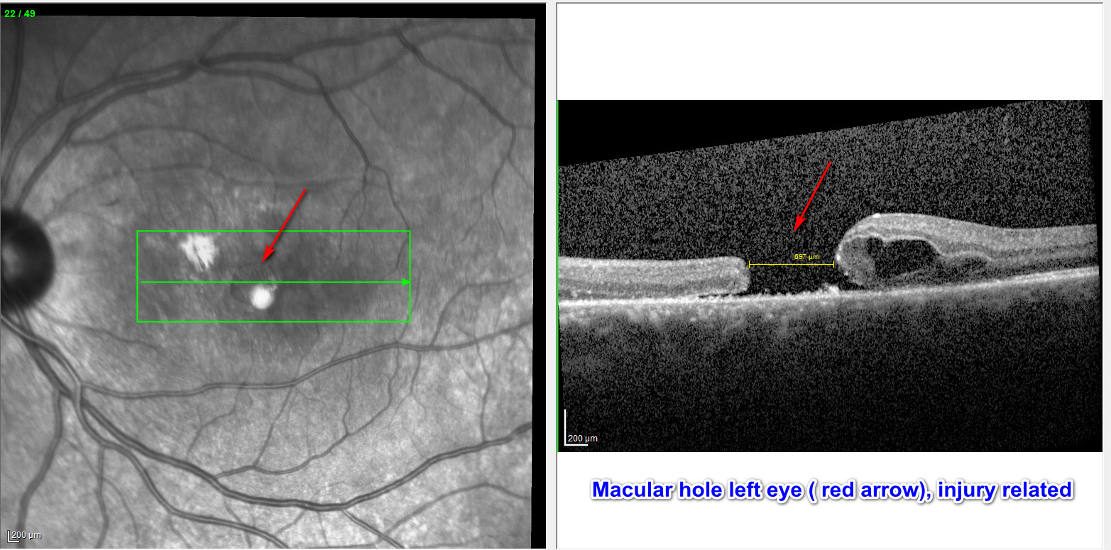
A macular hole is a full thickness defect at the center of the macula, the portion of the retina responsible for central vision. Imagine the film of a camera, where a hole is punched out at the center of the film. You can probably infer that the resulting picture would be clear in the periphery with a missing area in the center. With a macular hole, a blind spot is noted in the central field of vision. The peripheral vision is not affected by a macular hole except in rare events when a retinal detachment due to a hole occurs. A macular hole develops most often due to traction on the macula exerted by the vitreous, the gel which occupies the back compartment of the eye where the retina is located.
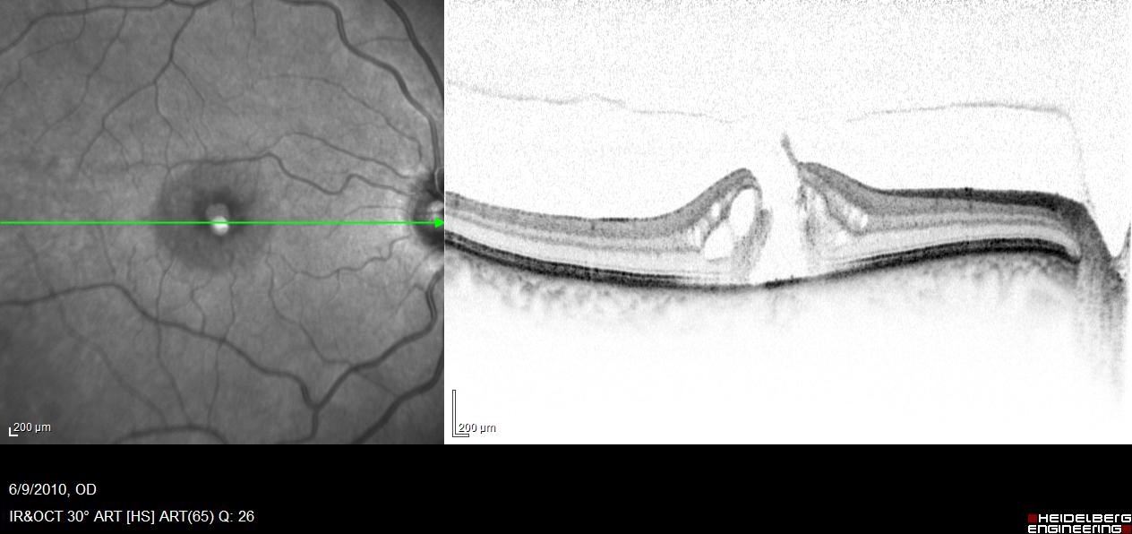
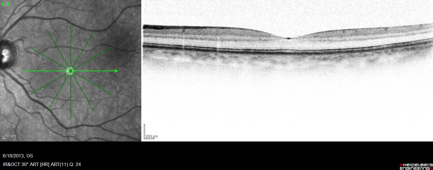
Photo of OCT Macula of normal eye and eye with hole
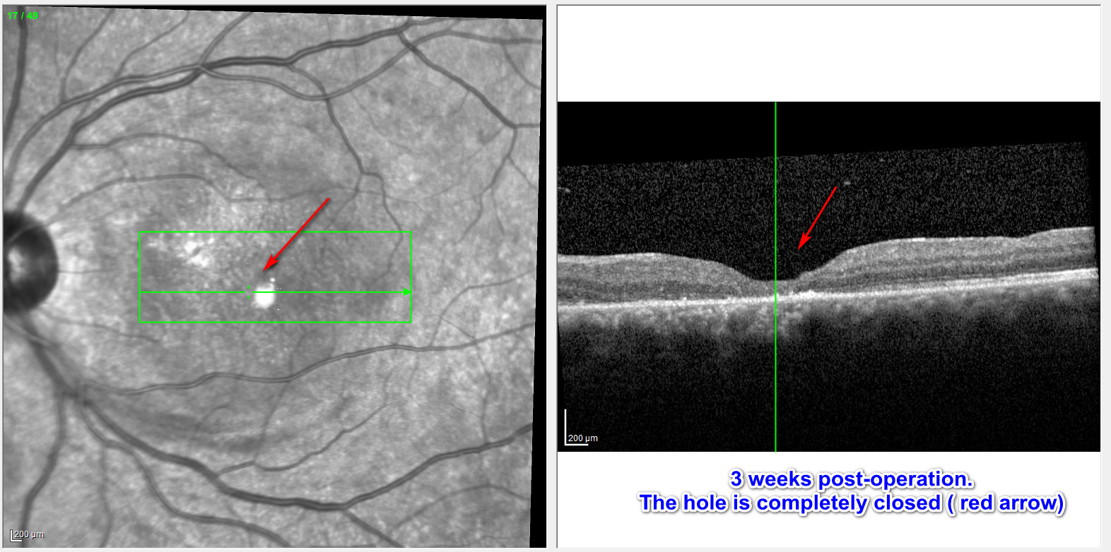
Treatment of a macular hole is usually a surgery called vitrectomy. With the surgery, the vitreous is removed and replaced with a temporary bubble of gas in the eye. Face down positioning is required after the surgery so that the gas bubble which floats upward may assist in pushing the edges of the hole together to seal it. In a few cases, an injection of medication called Jetrea could be considered as an alternative to vitrectomy. At the Retina Centers of Washington we have extensive experience with macular hole surgery and patients typically have good anatomical and visual outcomes.