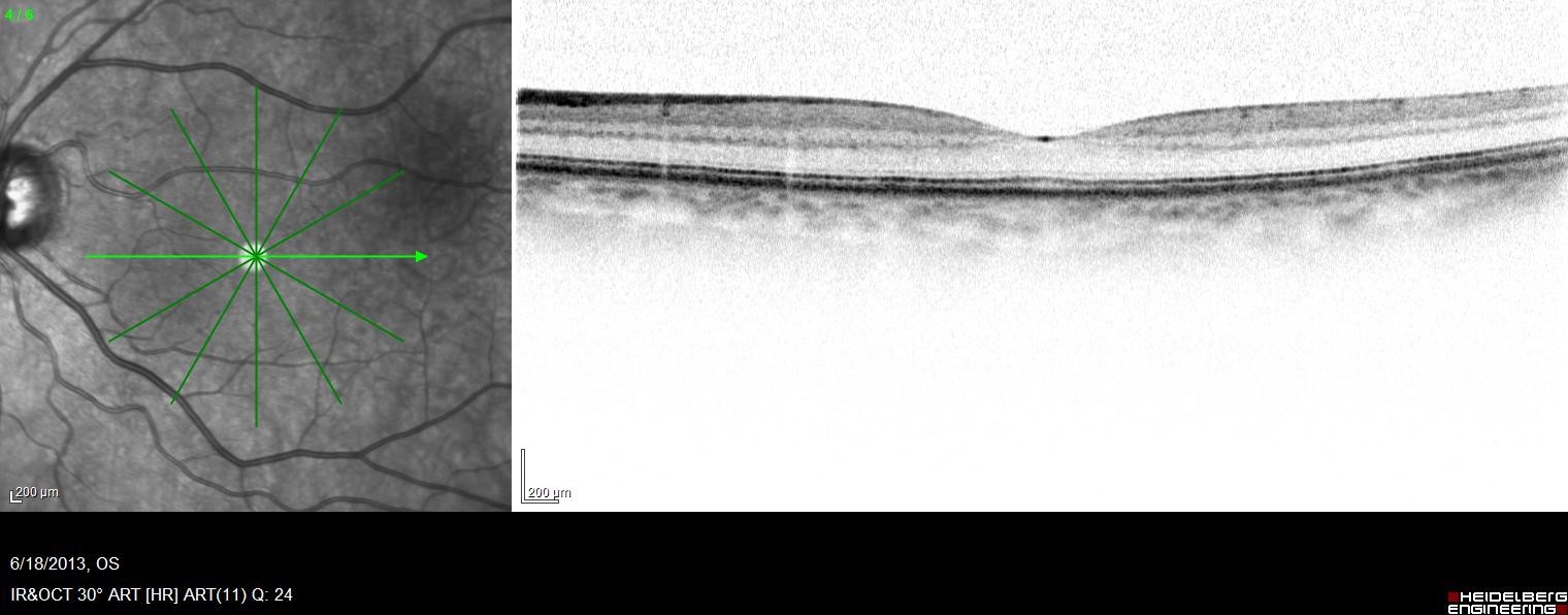
Macular puckering, also known as an epiretinal membrane, is the presence of a thin cellophane-like scar tissue over the surface of the macula, the portion of the retina that corresponds to an individual’s central field of vision. It forms most often from aging changes which occur to the vitreous, the gel which occupies the compartment of the eye where the retina is located. When the vitreous separates from the retina (please see the section on posterior vitreous detachment), most of the time the process occurs uneventfully. Sometimes, however, cells related to the vitreous may remain on the macula after the vitreous has separated. Over time, these cells can proliferate and form a macular pucker. This condition can also occur due to traumatic or inflammatory conditions of the eye. The presence of macular pucker may distort the contour of the macula.
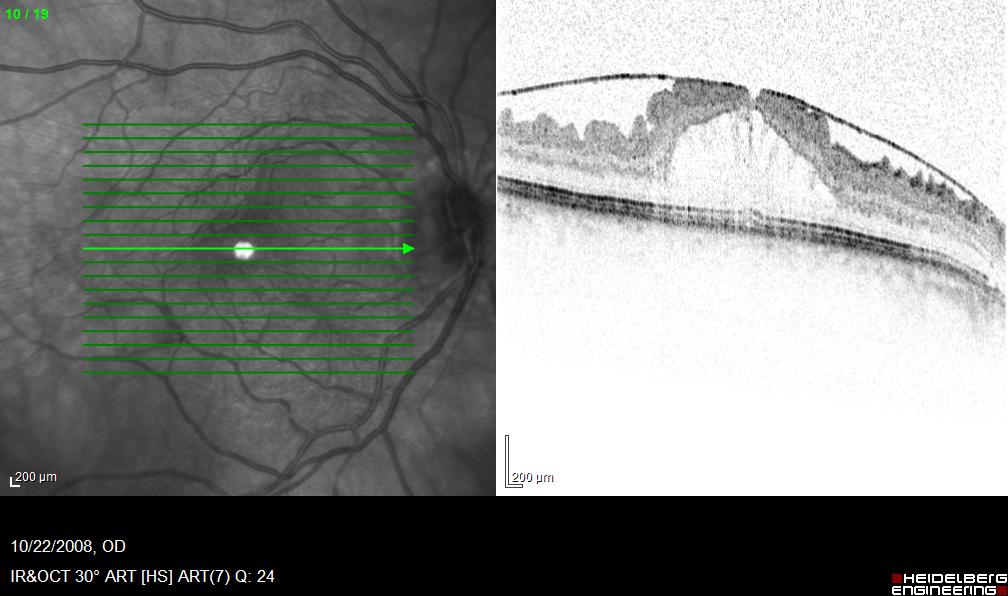
Side by side OCT photo of normal macula and ERM
In many circumstances, macular puckering does not significantly affect vision and may even be unnoticed to the individual affected. However, significant distortion of the contour of the macula may make the vision blurry or distorted. Some patients note metamorphopsia, where lines that are straight may look wavy to the affected eye.
The treatment of macular pucker consists of observation in a majority of cases. In more severe cases where significant blurring of vision or metamorphopsia develops and affects activities of daily living, a surgery called a vitrectomy is performed to improve the contour of the macula over time.