Your retina is supplied with oxygen and nutrients by blood vessels, arteries and veins. Arteries, which deliver blood to the retina, have a thicker wall. Think of it like a rubber garden hose. Veins, which deliver blood away from the retina, have a thinner and more collapsible wall.
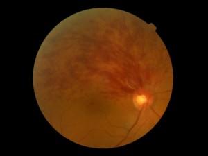
Picture of BRVO
A branch retinal vein occlusion (BRVO) occurs when a retinal artery compresses upon a retinal vein at the point where the two vessels may cross over each other. Flow of blood through the vein becomes more turbulent. Subsequently a clot forms thereby hindering the outflow of blood through the vein. Blood and clear fluid may build up around the distribution of the vein causing the retina in this region of the retina to become swollen. If the macula is affected, this is called macular edema. Sometimes retinal circulation may be permanently lost as well (capillary nonperfusion), which can lead to abnormal blood vessel growth (neovascularization).
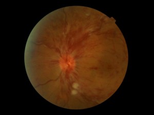
Picture of CRVO
This same process may also occur within the optic nerve where it connects to the rear of the eye. The central retinal artery (i.e. the main artery that feeds into and branches in the retina) and corresponding central retinal vein course through the optic nerve in close quarters. Similarly, the central retinal artery can cause a central retinal vein occlusion (CRVO). The loss of vision due to edema or neovascularization can sometimes be more severe with a CRVO, although this is highly variable.
Macular edema and neovascularization from retinal vein occlusions typically require treatment to stabilize and/or improve vision. Treatment consists of injections of medication and sometimes laser. If there is severe bleeding into the eye, surgery is sometimes performed for a vitreous hemorrhage.
At the Retina Centers of Washington we use anti-VEGF medications like Avastin and Eylea. Corticosteroid injections of Kenalog, Triescence, or Ozurdex are also used to manage macular edema from RVO. Generally earlier intervention from the time of presentation is associated with better visual outcomes, although the status of delivery of blood to the retina is also a critical factor.

